Retinoscopy is an optical diagnostic test used by eye care professionals to determine the refractive error of a patient’s eyes. It is a quick and accurate method of measuring the amount of correction required to produce a clear vision. The test is performed using a device called a retinoscope, which is an instrument that shines light into the eye to measure the reflection, also known as the “reflex.” This reflex helps the practitioner determine the power of lenses required to correct the patient’s vision.
Retinoscopy is an objective test, meaning that it does not rely on the patient’s input to determine the results. Retinoscopy is a useful tool for eye care professionals because it provides a fast and reliable method of determining the refractive error of a patient’s eyes. It is also a valuable tool for prescribing corrective lenses, as it helps practitioners determine the exact power of lenses required to correct the patient’s vision.
Principle of retinoscope:
Retinoscope works on Foucault’s principle. In the case of retinoscopy, a knife edge (peephole of retinoscope) placed on the principal axis of an optical system (patient eye reflex), intercepts a bundle of rays coming out of the patient’s eye. Depending on the position of the peephole of the retinoscope, various distributions of reflex can be observed in the reflex of the patient’s eye.
Optics behind retinoscope
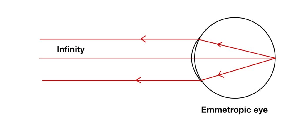
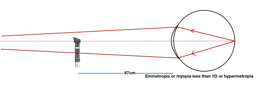
While doing retinoscopy on the patient we are less interested in rays which are going inside the eye, but we are more bothered about the ray which is exiting out of the eye. Suppose you are doing retinoscopy for an emmetropic eye from an infinite distance. In the case of emmetropes when rays enter the inside eye and rays focus on the retina. The rays which are reflected from the retina and which is also the focal point of the optical system of the eye in case of emmetropes, exits eye parallel. Suppose we now hold our retinoscope at infinity, and stop these rays, we will see a neutral glow.
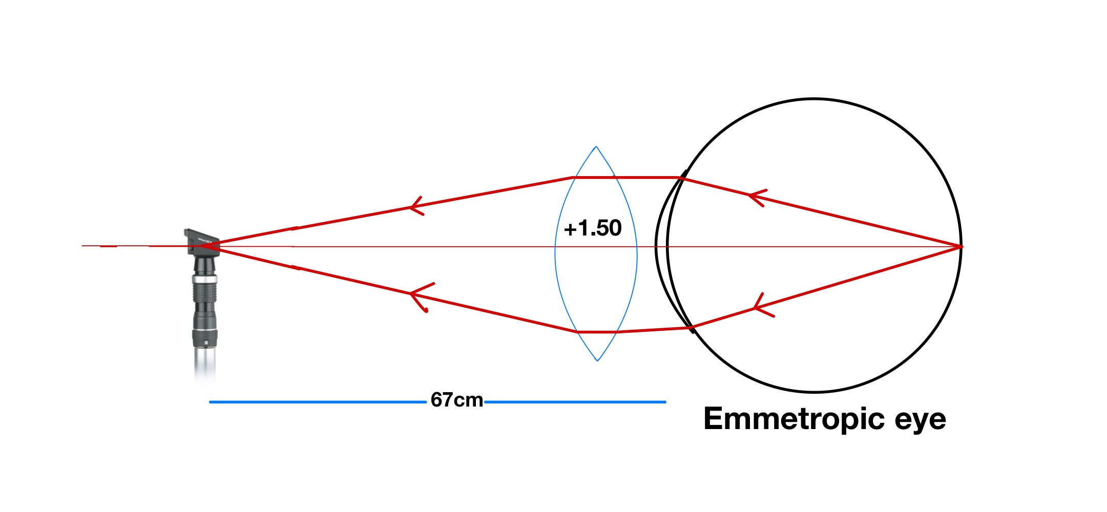
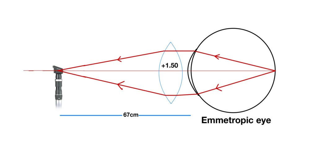
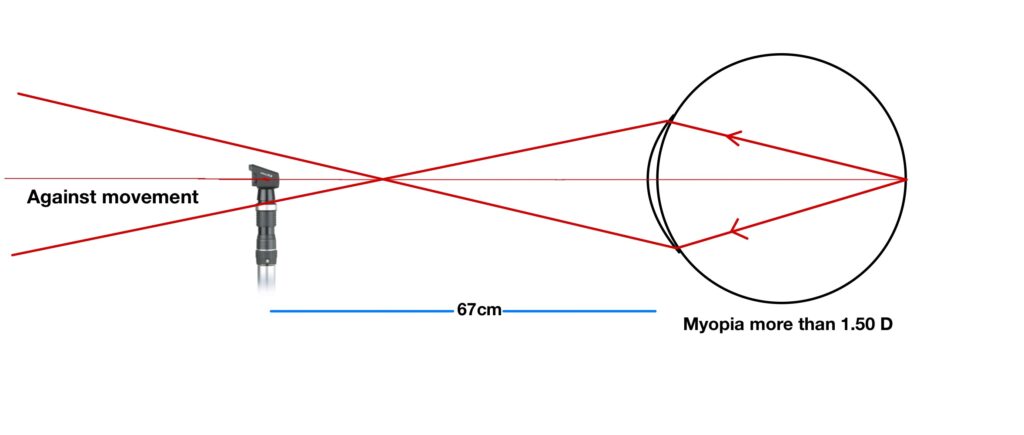
But it’s not practically possible to do retinoscopy from a distance which is situated at infinity. So, for the same reason, we come closer to the patient’s eye and use compensation for this decreased distance by using a plus lens in front of the patient’s eye. Simply if I put this, we know by now rays exit out of emmetropic eyes are parallel, so if you want to hold your retinoscope at your arm’s length (usually 67cm) far away from the patient’s eye and want to visualize the same reflex move which we saw at infinity. We have to use a lens which makes these parallel rays meet at the peephole of the retinoscope. In our case is only possible if we use +1.50 DS in front of the patient’s eye because the focal length of +1.50DS is approximately near 67cm.
If these rays, do not meet at the peephole of the retinoscope then we will see a move in reflex. Suppose rays meet somewhere behind the peephole retinoscope then we will see with movement and if front of the retinoscope we will see against movement.
Characteristics of retinoscopy reflex
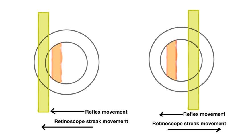
- The direction of movement of retinoscopic reflex:
Retinoscopic reflex moves with the movement of the retinoscope (i.e. when you sweep your retinoscope from left to right even reflex will also appear to move from left to right)
Retinoscopic reflex moves against with movement of retinoscope (i.e. when you sweep your retinoscope from left to right even reflex will appear moving against from right to left)
- Brightness: A dull glow will be seen when power is either high or in the case of media opacity. The reflex glow becomes bright as you approach towards neutrality
- Speed: The speed of glow is slow when you are far away from neutrality and become fast as you reach near to neutrality point.
- Thickness: Reflex becomes wider when it gets close to neutrality.
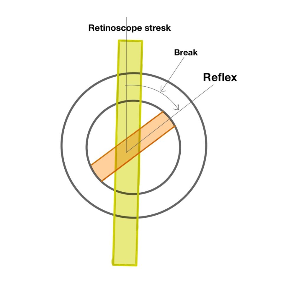
- Break: if your retinoscopy streak is not aligning with the reflex, we call it “break” which means the eye is having some astigmatism and the retinoscope streak is not aligning with any principal meridian of the eyes.
How to neutralize the reflex
The first step is to identify the type of refractive error (spherical or cylindrical)
Spherical
While seeing the reflex, start rotating your retinoscopy glow by rotating the retinoscope sleeve. If you see reflex is aligned with streak throughout while rotating retinoscope. Its mean refractive error is spherical.
Use plus lenses to neutralize with movement.
Use a minus lens to neutralize against movement.
Keep adding spherical power till the glow become neutral. Deduct compensation for hand distance from the power which you have used to find neutral glow, which will give you patient objective refraction.
Cylindrical
While seeing the reflex, start rotating your retinoscopy glow by rotating the retinoscope sleeve. If you see the reflex is not aligning with the streak throughout while rotating the retinoscope. Its mean refractive error is Cylindrical.
Find the principal meridian, which is done by rotating the sleeve of the retinoscope and finding a point where there is no break (i.e. retinoscope streak is aligning with the reflex).
See the movement of glow in that particular meridian. Remember movement retinoscope should be 90 away from the streak (i.e., if the streak is pointing towards 80 degree movement of the retinoscope, is given towards 170 degrees)
After this, you can choose two ways of neutralizing cylinder power
Method 1: Neutralization using spherical and cylindrical trial lenses:
Method 2: Neutralization using spherical trial lenses and an optical cross:
Method 1:
Step 1: Put the spherical trail lens that neutralizes one meridian (according to movement seen in step 1)
Step 2: Rotate your retinoscope streak 90° apart and neutralize the other principal meridian using a cylindrical lens. While placing the cylinder remember the cylindrical axis parallel to the retinoscope streak.
Step 3: Note down gross retinoscopic power from the trial frame
Step 4 Deduct +1.50 DS from gross retinoscopic spherical power to get net retinoscopic power.
Method 2:
Step 1: Put the spherical trail lens that neutralizes one meridian (according to movement seen). Note down the reading of spherical power and remove spherical power and streak orientation.
Step 2: Rotate your retinoscope streak 90° apart and neutralize the other principal meridian using spherical lenses. Note down the reading of spherical power and streak orientation.
Step 3: In this method, you will require a piece of paper and a pen. Draw an optical cross according to the orientation of the principal meridian. And put values in the optical cross, remember while putting value in the optical cross, values are recorded 90 degrees apart from streak orientation or the value will be written towards the direction of movement of the retinoscope.
Step 4: Deduct hand distance from each principal meridian and convert prescription from cross form to prescription, which gives the patient objective refraction value.
For more information about retinoscopy
Lecturer (Nethradhama School of Optometry)
Moptom