Types of retinoscopy includes
Static Retinoscopy
Dynamic retinoscopy
In static retinoscopy, the patient is instructed to focus on a distant target positioned 6 meters away, which is considered to be at optical infinity. The reason for using 6 meters as the standard distance for retinoscopy is that when light rays come from a source situated 6 meters away, they are typically parallel and do not stimulate accommodation. For example, if an emmetropic patient views an object located 1 meter away, they need to exert 1 dioptre (D) of accommodation to bring the rays into focus on the retina. If the object is 4 meters away, they require 0.25 D of accommodation for clear vision. However, when the object is 6 meters away, only 0.166 D of accommodation is needed, which is considered negligible. Static retinoscopy means retinoscopy is performed when accommodation is static or stable. Retinoscopy is performed to determine far point of the patient eye and working distance is deducted from gross retinoscopy value.
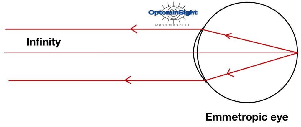
During retinoscopy, our main focus is on the ray of light that exits the eye rather than the rays entering it. When performing retinoscopy on an emmetropic eye from an infinite distance, we are interested in observing the behaviour of the rays that are reflected from the retina, which acts as the focal point of the eye’s optical system. In the case of emmetropes, these rays exiting the eye are parallel. If we were to hold our retinoscope at infinity and intercept these parallel rays, we would perceive a neutral glow.
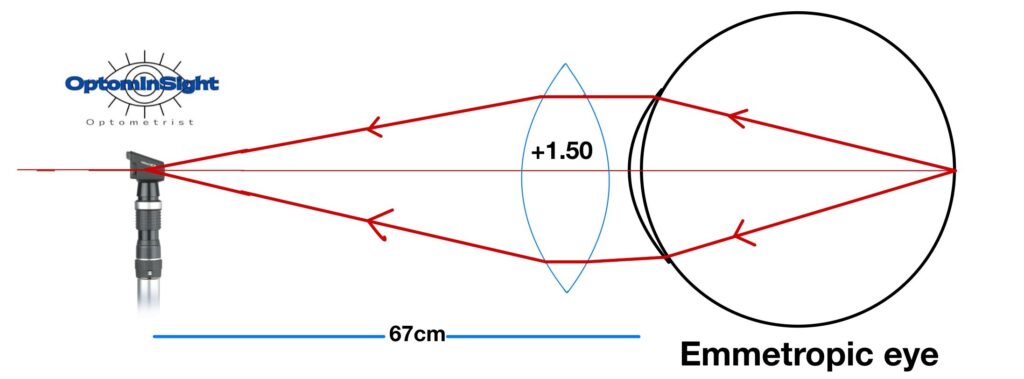
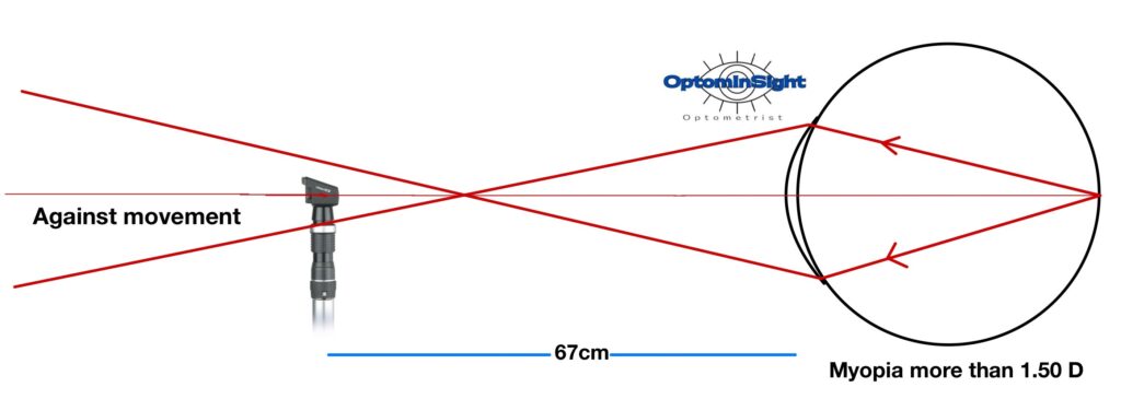
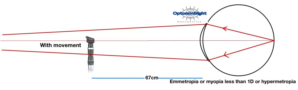
Due to the impracticality of performing retinoscopy from an infinite distance, we need to position ourselves closer to the patient’s eye. To compensate for this reduced distance, a plus lens is employed in front of the patient’s eye. In the case of emmetropic eyes, where the exiting rays are parallel, we aim to replicate the observation made at infinity by holding the retinoscope at a standard distance, typically around 67cm. To achieve this, we require a lens that converges the parallel rays and causes them to intersect at the peephole of the retinoscope. In this specific situation, a +1.50 dioptres sphere lens is utilized since its focal length is approximately 67cm, enabling the convergence of the rays at the desired distance.
If the parallel rays of light from the patient’s eye do not converge precisely at the peephole of the retinoscope, we will observe a movement in the reflex. If the convergence point of the rays is located behind the peephole, we will perceive with movement. Conversely, if the convergence point is in front of the retinoscope, we will observe against movement.
Read more about static retinoscopy by clicking link below
Dynamic retinoscopy
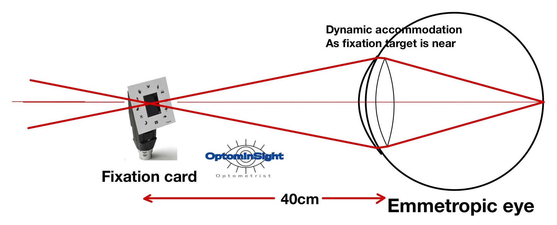
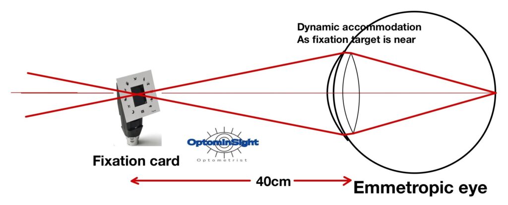
is a technique, when the patient’s accommodation is active rather than being at rest. During this procedure, the patient is instructed to focus on an object placed either near the retinoscope plane, behind the retinoscope, or at a position between the patient and the retinoscope. In dynamic retinoscopy, the usual deduction for working distance is not applied.
For instance, if a retinoscopy is performed at a distance of 40 cm, typically a deduction of +2 D would be made for the working distance. However, in dynamic retinoscopy, the subject is directed to fixate on a near target. In this case, let’s assume the subject is fixating on an object that is also located 40 cm away at the retinoscope plane. The subject will need to roughly accommodate an additional 2 D to see this target clearly. Therefore, if I perform dynamic retinoscopy on an emmetropic patient, I will observe a neutral point without placing any lens. The extra 2 D of accommodation is compensating for my working distance.
Types of dynamic retinoscopy
Lecturer (Nethradhama School of Optometry)
Moptom
👏👏👏