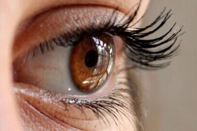
Many mechanisms exist to protect the eye from external injury.
1.Protective-mechanism-in-the-EyeMechanism of potential damages to the eye can ocular as:
- Mechanical
- Chemical
- Biological
- Electromagnetic radiation
1.Protection from mechanical damage
The orbit:
- The eye and orbital tissues are supported and shielded by the fat and bone walls of the orbit.
- The orbital fat acts as a semi-fluid padding that cushions the eye, providing shock absorption.
The eyelids:
- The eyelids act as a mechanical barrier between the eye and the outside world, quickly closing in response to reflexive or deliberate blinking.
- When triggered, the cilia (modified tiny hairs) on the eyelids’ skin provide a stimulus to blink in response to airborne particles.
The corneoscleral shell
- The corneoscleral shell gives the globe tensile strength.
- The cornea’s dense innervation enables quick blinking and withdrawal reflexes.
- Furthermore, corneal innervation supplies trophic elements that encourage epithelial recovery.
2.Protection from chemical damage
Eyelid closure
- Reflex blinking provides rapid closure of the eye in response to splash or foreign body sensation.
Bell’s phenomenon
- A normal Bell’s phenomenon provides involuntary upward and inward rotation of the globe on lid closure, removing the cornea from noxious stimuli .
Tears
- Tear flow increases in response to mechanical or noxious stimuli.
- This causes dilution and washout of the irritant.
Corneal epithelial barrier
- The corneal epithelium is 5-7 layers deep, with cells adjoined by desmosomes.
- Tight connections (zonulae occludens) surround the most superficial corneal epithelial cells, creating a low conductance barrier to fluid and solutes.
3.Protection from biological damage
Tear film and conjunctiva:
- Glycocalyx and mucous layer: A physical barrier against infections is provided by mucins in the glycocalyx (conjunctival cell membrane-bound mucin) and the mucous layer of the tear film, which can also trap bacteria.
- Aqueous layer: Many antibacterial substances, including as secretory immunoglobulin A (IgA), lysozyme, and lactoferrin, are present in the aqueous layer.
- Normal conjunctival flora: The normal bacterial flora may inhibit survival of more pathogenic species.
- Natural killer cells: Present in the conjunctiva, natural killer cells may have a role in restricting the spread of viral infection or tumors.
Corneal epithelium and Bowman’s layer:
- They serve as physical barriers to preventing microbiological infections.
Descemet’s membrane:
- In cases of severe corneal infections, Descemet’s membrane resists proteolysis, preserving the integrity of the globe.
4.Protection from electromagnetic radiation
Eyelid closure:
- The dazzle reflex causes reflexive blinking in response to bright light.
Pupil constriction:
- Rapid pupil constriction in response to bright light limits excessive radiation exposure to the ocular media internal to the iris.
Light absorption by ocular tissues:
- The cornea and sclera absorb ultraviolet (UV)-B, UV-C, infrared (IR)-B, and IR-C .
- The crystalline lens absorbs UV-A.
- Excessive UV-induced oxidative damage is prevented by antioxidants in the lens and macula.
- Short-wavelength light is absorbed by the yellow macular carotenoid xanthophyll pigments in Henle’s fibre layer.
- Melanin and hemoglobin, which are primarily present in the choroid, absorb too much IR radiation and light.

